Introduction
Keratoconus and LASIK are two terms that often come up in discussions about vision correction and eye health. Understanding the relationship between these conditions and the potential risks involved is crucial for anyone considering corrective eye surgery. This article aims to provide a comprehensive overview of keratoconus, corneal ectasia, their symptoms, and the available treatments. We will also explore the risks associated with LASIK for keratoconus patients and discuss safer alternatives for vision correction.
What Is Keratoconus?
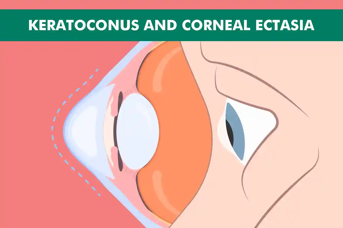
Keratoconus is a progressive eye disease where the cornea, which is normally round, thins out and begins to bulge into a cone-like shape. This can cause significant visual impairment as the cornea’s irregular shape prevents light from focusing correctly on the retina.
Common symptoms of keratoconus include:
- Blurred or distorted vision
- Difficulty with everyday tasks such as reading, driving, and recognizing faces
- Increased sensitivity to light (photophobia), causing discomfort in bright environments
- Frequent changes in eyeglass prescriptions due to the progressive nature of the disease altering the shape of the cornea
The exact cause of keratoconus is not known, but it is believed to be a combination of:
- Genetic factors (e.g., family history of the condition)
- Environmental influences (e.g., chronic eye rubbing, exposure to ultraviolet rays)
- Hormonal changes affecting the structural integrity of the cornea
What Is Corneal Ectasia?
Corneal ectasia is a condition that can develop after refractive surgery like LASIK. It is characterized by a progressive thinning and bulging of the cornea, similar to keratoconus, but typically occurs as a complication rather than a primary condition.
Symptoms of corneal ectasia include:
- Worsening vision
- Gradual decline in visual clarity and sharpness
- Difficulty performing everyday tasks such as reading or driving
- Ghosting or double images
- Objects appear to have a shadow or duplicate
- Complicates visual perception
- Increased sensitivity to light (photophobia)
- Significant discomfort in bright environments
- Challenges being outdoors or in well-lit areas
Diagnosis usually involves:
- Corneal topography
- Sophisticated imaging technique
- Maps the shape of the cornea
- Measures corneal thickness
- Provides detailed information about corneal structure and abnormalities
- Crucial for identifying the extent of corneal ectasia
- Helps in planning appropriate treatment strategies
LASIK and Keratoconus: Understanding the Risks
Given the structural instability of the cornea in keratoconus patients, LASIK is generally unsafe. The procedure can worsen the compromised corneal structure, leading to complications like corneal ectasia, which severely impairs vision and quality of life.
Safer alternatives include Corneal Cross-Linking (CXL) and specialized contact lenses. CXL strengthens the cornea by increasing collagen cross-links, halting keratoconus progression. Specialized contact lenses, such as scleral or hybrid lenses, improve vision by creating a smooth refractive surface, offering comfort and stability.
What Is Corneal Cross-Linking (CXL)?
Corneal Cross-Linking (CXL) is a minimally invasive procedure designed to strengthen the cornea and halt the progression of keratoconus. The treatment involves applying riboflavin (vitamin B2) eye drops to the cornea, which is then activated by ultraviolet (UV) light.
CXL works by increasing the number of collagen cross-links in the cornea, thereby making it more rigid and less likely to bulge. This procedure can be a viable option for patients with early to moderate keratoconus.
Other Vision Correction Options for Keratoconus Patients
1. Contact Lenses:
Soft Contact Lenses: Initially used for mild keratoconus, these lenses provide comfort but may not correct vision as effectively in advanced stages.
Rigid Gas Permeable (RGP) Lenses: These lenses offer better vision correction by creating a smooth refractive surface over the irregular cornea.
Hybrid Lenses: Combining a hard center with a soft outer ring, these lenses provide the clarity of RGP lenses with the comfort of soft lenses.
Scleral Lenses: Larger lenses that vault over the cornea and rest on the sclera, providing excellent vision correction and comfort for advanced keratoconus.
2. Corneal Cross-Linking (CXL):
Standard CXL: Involves applying riboflavin (vitamin B2) to the cornea and then exposing it to ultraviolet light, strengthening the corneal tissue to halt progression.
Accelerated CXL: A faster version of the standard procedure, using higher intensity UV light for a shorter duration, reducing treatment time.
3. Intacs:
Intrastromal Corneal Ring Segments (ICRS): Small, crescent-shaped plastic inserts placed in the cornea to flatten it and improve vision. This minimally invasive procedure can delay the need for a corneal transplant.
4. Topography-Guided Custom Ablation:
Laser Treatment: Uses detailed corneal maps to guide a laser in reshaping the cornea, improving vision, and reducing irregularities. Often combined with CXL for better outcomes.
5. Phakic Intraocular Lenses (IOLs):
Implantable Lenses: These lenses are placed inside the eye, in front of the natural lens, to correct refractive errors. Suitable for patients who cannot achieve good vision with contact lenses or glasses.
6. Corneal Transplant:
Penetrating Keratoplasty (PK): Full-thickness corneal transplant, replacing the entire cornea with a donor cornea. Used for severe keratoconus cases.
Deep Anterior Lamellar Keratoplasty (DALK): Partial-thickness transplant, replacing only the front layers of the cornea, preserving the patient’s own endothelial cells and reducing rejection risk.
Preventing Corneal Ectasia
Preventing corneal ectasia in refractive surgery involves careful preoperative assessments and ongoing monitoring to minimize risks.
1. Preoperative Screening
Advanced imaging tools, like corneal topography, assess corneal thickness and shape to identify risk factors such as keratoconus, helping to avoid surgery in high-risk patients.
2. Procedure Selection
For patients with thinner corneas, alternative procedures like SMILE or PRK, which don’t require a corneal flap, reduce the risk of ectasia.
3. Limiting Tissue Removal
Surgeons must avoid removing too much corneal tissue, ensuring sufficient residual stromal thickness to maintain corneal strength.
4. Postoperative Monitoring
Regular follow-up helps detect early signs of ectasia. Early intervention, such as corneal cross-linking, can prevent progression.
5. Corneal Cross-Linking (CXL)
CXL strengthens the cornea by increasing collagen cross-links, offering protection against ectasia for high-risk patients or those with early symptoms.
With these measures, the risk of corneal ectasia can be significantly reduced, ensuring safer refractive surgeries.
FAQs
1. Can I have LASIK if I have mild keratoconus?
LASIK is not recommended for keratoconus, even if mild, as it can worsen the condition. Consider alternative treatments.
2. How do I know if I have corneal ectasia?
Corneal ectasia is diagnosed through an eye exam with corneal topography. Symptoms include blurred vision, double vision, ghosting, and light sensitivity. Consult an eye care professional if you experience these.
3. Is corneal cross-linking a permanent solution for keratoconus?
Corneal cross-linking stabilizes keratoconus but is not a cure. Regular follow-ups are needed to monitor stability.
4. What are the best alternatives to LASIK for keratoconus patients?
Alternatives include corneal cross-linking, scleral contact lenses, Intacs, and in advanced cases, corneal transplant. Discuss options with your eye care provider.
5. Can corneal ectasia be reversed?
Corneal ectasia cannot be reversed but can be managed with treatments like corneal cross-linking. Early detection and treatment are key to maintaining vision.

